The group has increasingly got involved in local probe microscopies, especially after the installation of a new cryogenic microscope. A closed-cycle helium gas, high vector field microscopy equipment (Attocube AttoDry1000 MFM-SHPM) has been recently improved with a second cryogenic insert for Hall bar microscopy. With this equipment, thus, two magnetic microscopes can operate on samples at a temperature down to 4 K in separate cryogenic inserts: (1) a non-invasive probe, scanning Hall probe microscope (SHPM) and (2) a force microscope (MFM, AFM) with interferometric deflection detection. Analysis of magnetic images can be assisted by micromagnetic modelling. Another two atomic force microscopes are available for room-temperature studies under in-situ magnetic field, for electrical conductance microscopy and for inspection purposes. One Solver MFM (NDT-Europe) microscope is equipped with an electromagnet to provide magnetic field up to 2 kG in the sample-probe area. For inspection purposes a table-top Nanosurf AFM ready for operation in a few modes such as contact or taping AFM, MFM and conductive tip ct-AFM.
![]()
![]()

Left panel is a MFM phase contrast image of Permalloy film deposited on a triangular silicon grating, measured at remanence after saturation in the Grating Axis ( ) - hard magnetic axis -. Right panel is sketch of the micromagnetic domain distribution of the antiparallel domains formed at transverse remanence (Nanoscale, 2025).

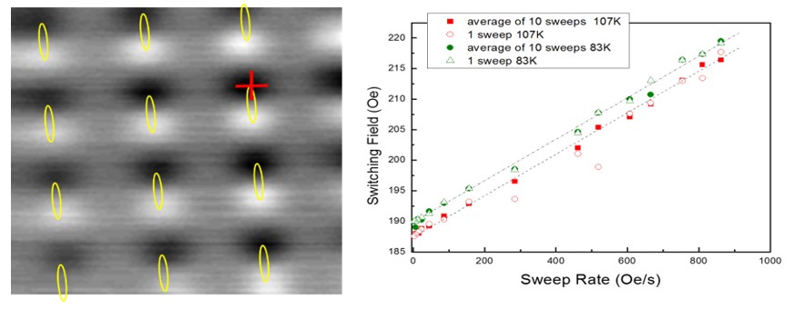
Left panel is Hall effect (SHPM) image scanned above an array of 20 nm thick permalloy microellipses after application of a saturating field along the ellipse major axis. Each of these ellipses makes a dipole fingerprint of single-domain with easy axis magnetization. Image size 20×20 µm2. Drawn for clarity are yellow lines corresponding to microellipse borders as obtained separately from SEM. Red cross points the position of Hall probe for dynamic study of the easy axis reversal: right panel plots the switching field measured for different field rates, number of sweeps or temperature (see legend).

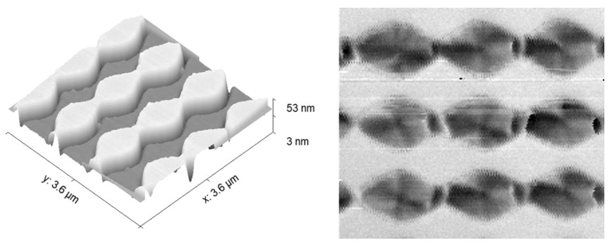
Force images of an array of microscale permalloy pseudo-wires measured in dual-pass dynamic mode with a magnetic tip: Left panel is the topography image obtained with the first pass and right panel is the magnetic force image (phase shift) as obtained from the second pass at a fixed distance above the surface defined by the first pass. Image phase spans 8.5 deg. Sample fabricated by Alvaro Muñoz-Noval (UCM) and measured with the Attocube MFM microscope.

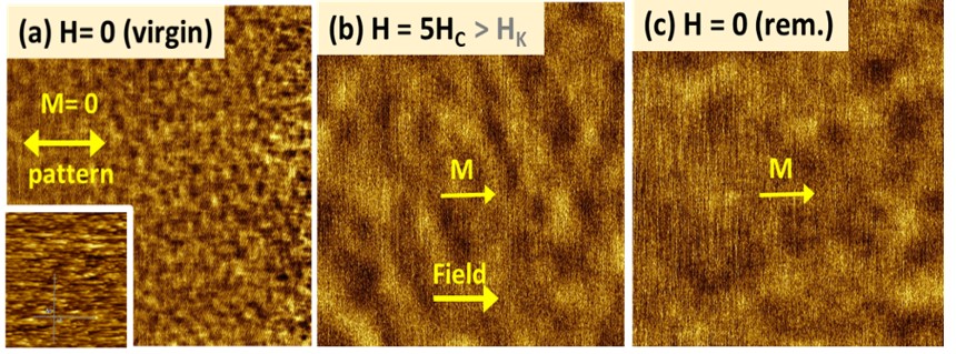
Magnetic Force Microscopy 6 x 6 mm2 scans of an IBS patterned cobalt film in easy axis magnetization i.e. with field applied along the ripple pattern. (a) Virgin state before the application of any magnetic field (dipole-like elements separated by ~ 0,3 mm). Inset shows the ripple pattern surface of AFM image 2 x 2 mm2 (b) Saturation state with in-situ applied field H = 100 mT – contrast due to magnetization ripple -, and (c) Remanence state after the removal of field. (J. Phys. D: Appl. Phys. 2016)

Left panel is dynamic mode AFM image of a permalloy film surface deposited on a LIPSS polymer foil (LIPSS means Laser Induced Patterning of Surface Structures). Image size is 10 x 10 µm2. This thin permalloy film (thickness up to at least 30 nm) is a nanoscale very corrugated, continuous magnetic system whereby the hard axis magnetization perfectly follows the surface contour (undulated state): the right panel shows volume-like magnetic poles in such magnetic profile as calculated with micromagnetic modelling and fitting Moke loops (J. Magn. Magn. Mater. 2020)